
Best Guide to Spine Surgery Instruments & Craniotomy Tools (2026)
August 6, 2026 2026-08-06 10:52Best Guide to Spine Surgery Instruments & Craniotomy Tools (2026)
 hasni
hasni 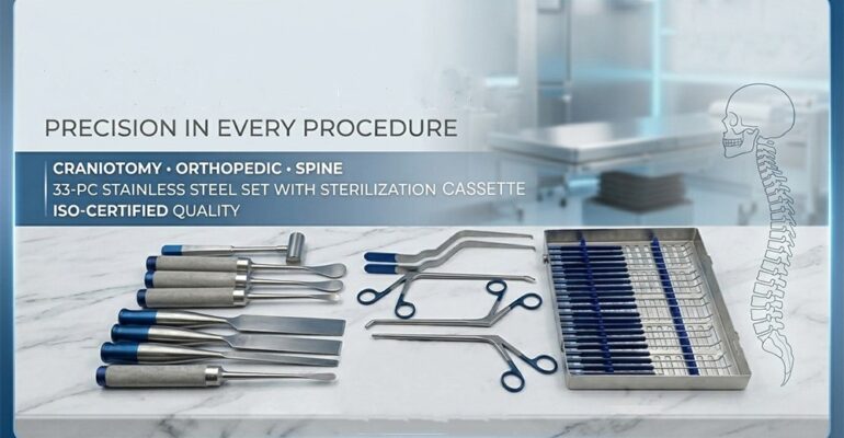
Best Guide to Spine Surgery Instruments & Craniotomy Tools (2026)
Two Specialties, One Shared Engineering Challenge
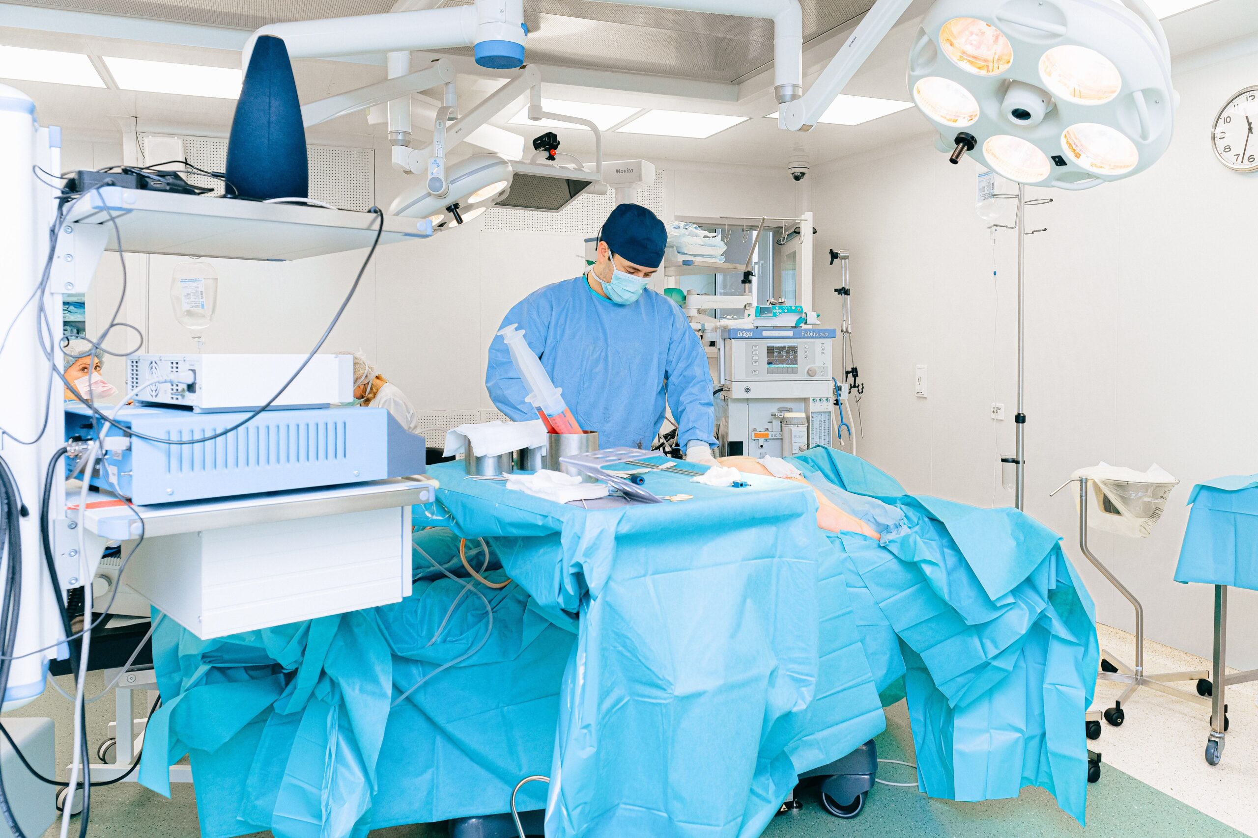
Spine surgery and craniotomy sit at opposite ends of the body, but they share a core engineering challenge: gaining safe, controlled access to bone that directly protects the central nervous system, without damaging the neural tissue underneath. Whether it’s a surgeon removing a lamina to decompress a nerve root in the lumbar spine, or a neurosurgeon cutting a bone flap to access the brain, the instruments involved have to combine cutting precision with built-in protection for the tissue just beneath the bone. This guide covers the essential instrument categories for both spine surgery and craniotomy, what each tool actually does, and what to look for when building or sourcing a tray for either specialty in 2026.
Part 1: Spine Surgery Instruments
Pedicle Screw Systems
Pedicle screw systems remain the backbone of modern spinal fixation surgery, composed of screws, rods, and connectors that provide rigid stabilization of the vertebral column in cases of trauma, tumor, or degenerative instability. Screws are placed through the vertebral pedicles and secured to rods spanning the affected levels, distributing mechanical load and supporting bone fusion between vertebrae. Our dedicated Spinal Fixation Instruments guide covers screw types, rod systems, and cage options in more depth.

Laminectomy and Decompression Instruments
Laminectomy — removing a portion of the vertebral lamina to relieve pressure on nerve roots or the spinal cord — relies on a specific instrument set: Kerrison rongeurs for controlled bone removal at the canal margin, laminectomy curettes for scraping and shaping bone, high-speed surgical drills for thinning the lamina before final removal, and spinal retractors to maintain a clear working field throughout. Kerrison rongeurs come in different footplate thicknesses (commonly 1mm or 2mm) specifically so surgeons can complete decompression with a thinner, safer footprint near the dura.
Microdiscectomy and Minimally Invasive Instruments
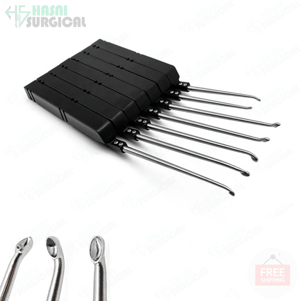
Microdiscectomy — removing herniated disc material compressing a nerve root — is typically performed through a small incision using specialized micro-instruments under magnification. A dedicated microdiscectomy set typically includes fine pituitary rongeurs, nerve root retractors, micro-curettes, and a narrow-bladed knife for disc annulus incision, all sized for work through a limited surgical corridor.
Cervical and Lumbar-Specific Instrumentation
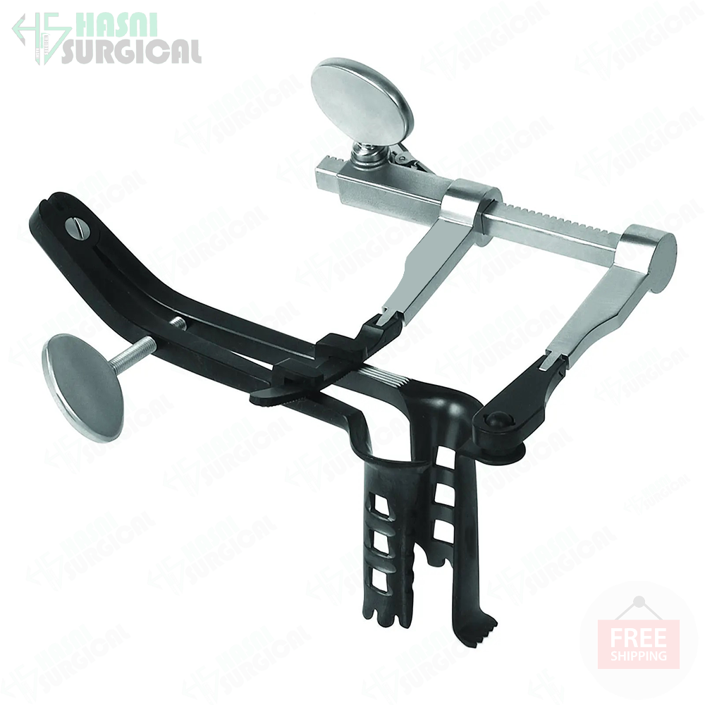
Cervical spine surgery — whether anterior or posterior approach — typically uses a dedicated instrument set distinct from lumbar instrumentation, given the smaller working corridor and proximity to critical vascular and neural structures in the neck. A Caspar-style micro lumbar retractor system, for example, provides distraction and access specifically suited to anterior lumbar interbody procedures, while cervical sets are scaled down accordingly for the smaller vertebral anatomy of the neck.
Spinal Probes and Neural Monitoring Instruments
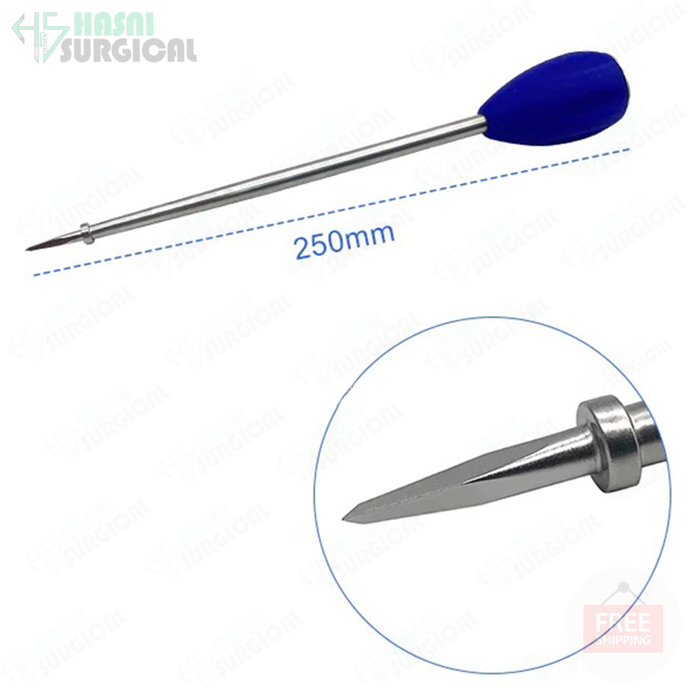
Ball-tip and angled spinal probes are used throughout decompression and fusion procedures to palpate the pedicle wall, confirm screw trajectory integrity, and check for breach before rod placement — a low-tech but essential safety check that remains standard practice even in image-guided and navigation-assisted spine surgery.
Part 2: Craniotomy Instruments
What a Craniotomy Actually Involves
A craniotomy is the surgical removal of a section of the skull — the bone flap — to access the brain for tumor resection, hematoma evacuation, aneurysm repair, or other neurosurgical procedures, after which the bone flap is typically replaced and secured with plates and screws. According to a detailed overview on craniotomy published on NCBI’s StatPearls, the procedure requires a full interprofessional surgical team and a specific, well-defined instrument tray to be carried out safely.
The Core Craniotomy Instrument Tray
Per the StatPearls overview, a standard craniotomy tray includes: a periosteal elevator for reflecting the scalp and separating tissue from bone; bayonet and Adson forceps for tissue handling; a head-fixation system to immobilize the skull during bone work; a high-speed pneumatic cranial drill (craniotome) with perforating bits; a round burr attachment; a Gigli wire saw with guide and handles for completing the bone cut between burr holes; a bone curette; a Kerrison bone rongeur; a Penfield dissector; Gerald forceps; and hemostatic agents such as bone wax, Surgicel, and Gelfoam.
A detailed technique description from Medscape’s craniotomy technique reference walks through how these instruments are used in sequence: burr holes are drilled with a perforator tip, then widened with a curette and Kerrison rongeur once the dura is carefully exposed. Once the full bone flap outline is drilled, a flap elevator lifts the bone while a Penfield No. 3 dissector separates the underlying dura, after which the flap is removed and set aside to be replated at closure.
Why the Gigli Saw Still Has a Place in Craniotomy
Even with modern high-speed pneumatic craniotomes widely available, the Gigli wire saw remains part of the standard craniotomy tray specifically because it can be threaded between two burr holes and drawn through the bone without needing the same clearance a rigid powered blade requires — useful in specific anatomical situations or as a reliable backup when power equipment isn’t an option. Our full Gigli saw guide covers its history, technique, and care in more detail.
Protecting the Dura: The Most Delicate Step
Once the bone flap is removed, dural elevators and dissectors are used to carefully separate and protect the dura mater — the membrane covering the brain — from the inner surface of the skull. Proper handling at this stage is critical for preventing cerebrospinal fluid leaks and other post-operative complications. Retractors, including self-retaining systems, are then used to gently hold back brain tissue and maintain a clear surgical field, with brain spatulas and cottonoids protecting tissue from direct pressure throughout the procedure.
Hemostasis During Cranial Bone Work
Bleeding from bony edges during a craniotomy is typically controlled with bone wax, while epidural bleeding — which tends to be more diffuse — is managed with hemostatic agents, Gelfoam, and thrombin-soaked cottonoids. Major vessels such as the middle meningeal artery are controlled with bipolar cautery before the dural opening proceeds.
Where Spine and Cranial Instrument Trays Overlap
- Rongeurs: Kerrison rongeurs appear in both spine decompression and craniotomy trays, differing mainly in footplate size and angle suited to each anatomical region.
- Bone curettes: Used for shaping and smoothing bone edges in both lumbar laminectomy and cranial bone flap procedures.
- Wire saws: The Gigli saw crosses over from craniotomy into orthopedic and spinal deformity correction procedures requiring manual bone cutting in confined spaces.
- Hemostatic agents: Bone wax and Gelfoam are standard across both specialties for controlling bleeding from cut bone surfaces.
- Retractor systems: Self-retaining retractors are essential in both fields for maintaining a stable surgical field without requiring constant manual holding.
What to Look for When Sourcing These Instrument Sets
- Certification: ISO 13485 and CE marking are standard expectations for neurosurgical and spinal instrument manufacturers exporting internationally.
- Material grade: Surgical-grade stainless steel with proper heat treatment for cutting-edge instruments like rongeurs and curettes, which need to hold precision edges through repeated sterilization.
- Complete set configuration: Confirm the tray includes the full range needed for your specific procedure type — a lumbar decompression set and a cervical set are not interchangeable.
- Sterilization compatibility: ISO 17664/11607-2 documentation for validated reprocessing, particularly important for delicate micro-instruments used in microdiscectomy and craniotomy.
Hasni Surgical manufactures spine and neurosurgical instrumentation from its Sialkot, Pakistan facility, supplying hospitals and distributors internationally. Reach the team by phone at +92 308 5391894, by email at info@hasnisurgical.com, or through hasnisurgical.com.
Related Reading
For a deeper look at spinal fixation hardware specifically, see our Spinal Fixation Instruments guide and Professional Spine Stabilization Tools guide. For manual bone-cutting technique specifically relevant to craniotomy, see our Gigli Saw guide. Our overview of how surgical instruments are sterilized also applies directly to reprocessing these instrument trays between cases.
Frequently Asked Questions
Q1: What instruments are included in a standard craniotomy tray?
A standard tray includes a periosteal elevator, bayonet and Adson forceps, a head-fixation system, a high-speed pneumatic craniotome with perforating bits, a Gigli wire saw with guide and handles, a bone curette, a Kerrison rongeur, a Penfield dissector, Gerald forceps, and hemostatic agents like bone wax and Gelfoam.
Q2: What is a Kerrison rongeur used for?
A Kerrison rongeur is used to remove bone in a controlled manner at the margins of a laminectomy or craniotomy bone opening, available in different footplate thicknesses to allow progressively finer, safer bone removal near the dura or spinal canal.
Q3: Is the Gigli saw still used in modern craniotomy?
Yes. Despite the availability of high-speed pneumatic craniotomes, the Gigli wire saw remains part of the standard craniotomy tray because it can be threaded between burr holes and drawn through bone without needing the clearance a rigid powered blade requires.
Q4: What is the difference between a laminectomy set and a microdiscectomy set?
A laminectomy set is built around larger instruments like Kerrison rongeurs and high-speed drills for open decompression, while a microdiscectomy set uses finer, smaller instruments such as pituitary rongeurs and micro-curettes designed for work through a limited surgical corridor under magnification.
Q5: Why are dural elevators considered such a critical instrument in craniotomy?
Dural elevators are used to carefully separate the dura mater from the inner skull surface, and improper handling at this stage can lead to cerebrospinal fluid leaks and other serious post-operative complications, making precision instrument quality especially important here.
Q6: How is bleeding controlled during bone work in spine and cranial surgery?
Bone wax controls bleeding directly from cut bone edges, while hemostatic agents, Gelfoam, and thrombin-soaked cottonoids manage more diffuse bleeding such as epidural bleeding during craniotomy, with bipolar cautery used to control larger vessels.
Q7: What is a pedicle screw system used for?
Pedicle screw systems provide rigid internal fixation of the vertebral column, composed of screws placed through the vertebral pedicles connected by rods, used to stabilize the spine in cases of trauma, tumor, or degenerative instability and to support bone fusion.
Q8: What certifications should I look for when sourcing spine or craniotomy instrument sets?
At minimum, confirm ISO 13485 certification and CE marking for the European market, along with ISO 17664/11607-2 documentation for validated reprocessing and sterile packaging, particularly important for delicate micro-instruments.
Q9: Why does cervical spine instrumentation differ from lumbar instrumentation?
The cervical spine has a smaller working corridor and sits close to critical vascular and neural structures in the neck, so cervical instrument sets are scaled down and configured differently compared to lumbar sets built for a larger working space.
Q10: What role do spinal probes play during pedicle screw placement?
Ball-tip and angled spinal probes are used to palpate the pedicle wall and confirm screw trajectory integrity before rod placement, serving as a manual safety check even in image-guided and navigation-assisted spine surgery.
Conclusion
Spine surgery and craniotomy each demand highly specific instrument configurations, but both fields share the same underlying engineering principle: giving surgeons controlled access to bone while actively protecting the neural tissue directly beneath it. From pedicle screw systems and Kerrison rongeurs in spinal decompression, to craniotomes, Gigli saws, and dural elevators in cranial access, the quality and precision of these instruments directly affects surgical safety and patient outcomes. Whether you’re building a spine surgery tray, a craniotomy set, or both, verifying certification, material grade, and sterilization compatibility should be non-negotiable steps before sourcing.
