
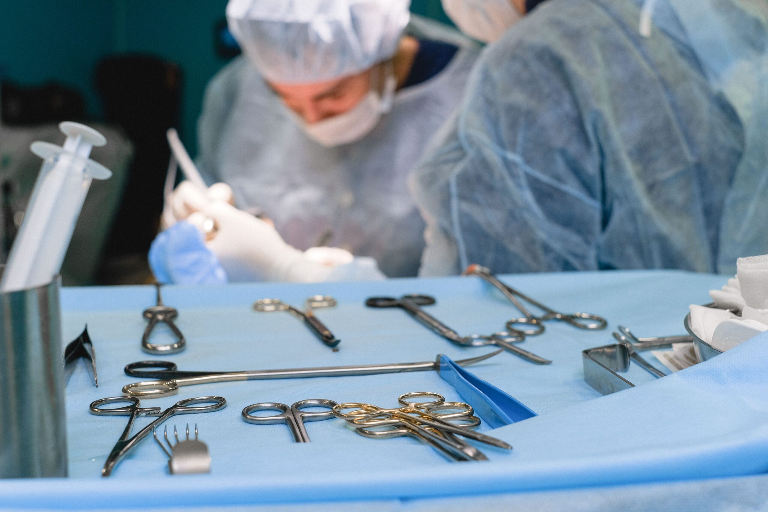
Professional Spine Stabilization Tools: Advanced Instruments for Safe Spinal Surgery
June 23, 2026 2026-06-23 5:48Professional Spine Stabilization Tools: Advanced Instruments for Safe Spinal Surgery
Professional Spine Stabilization Tools: Advanced Instruments for Safe Spinal Surgery
Spinal instability is one of the most debilitating conditions a patient can experience. Whether caused by degenerative disc disease, vertebral fracture, scoliosis, spondylolisthesis, tumor, or infection — an unstable spine compresses nerve roots, damages the spinal cord, causes severe chronic pain, and progressively destroys a patient’s quality of life. When conservative management fails, spinal stabilization surgery is the definitive solution.
But spinal stabilization surgery is only as safe and effective as the tools used to perform it. The confined surgical corridor of the spine, the proximity of the spinal cord and major vessels, and the extraordinary mechanical demands of fixation constructs that must last decades — all of these factors demand professional spine stabilization tools of the highest quality, precision, and reliability.
This advanced guide covers the full spectrum of professional spine stabilization instruments — from the tools used for safe pedicle access to advanced deformity reduction systems, interbody fusion instrumentation, and the latest minimally invasive stabilization tools — along with clinical selection criteria and quality standards for procurement teams and spine surgery programs worldwide.
“Spinal stabilization is not a procedure for improvisation. Every instrument placed near the spinal cord must perform flawlessly — there is no margin for tool failure in spine surgery.”
Understanding Spinal Instability: Why Stabilization Surgery Is Needed
The spine achieves its remarkable combination of strength, flexibility, and protective function through the coordinated action of its bony elements, intervertebral discs, ligaments, and muscles. When any of these components are compromised — by degeneration, injury, disease, or surgical disruption — spinal instability results.
Spinal instability is defined as the loss of the spine’s ability to maintain its normal pattern of displacement under physiological loads without initial or additional neurological deficit, major deformity, or incapacitating pain. The most common causes requiring surgical stabilization include:
- Degenerative disc disease and spondylosis: Progressive disc height loss and facet degeneration destabilize the motion segment, causing painful micromotion and nerve root compression
- Spondylolisthesis: Forward slippage of one vertebra on the next — causing back pain, leg pain, and progressive neurological deficit in higher-grade slips
- Vertebral fractures: Burst fractures, flexion-distraction injuries, and fracture-dislocations disrupt the three-column stability of the spine, often with associated cord injury
- Spinal tumors: Primary vertebral tumors and metastatic disease destroy bone, cause pathological fractures, and compress neural elements
- Spinal infections: Pyogenic discitis/osteomyelitis and tuberculosis (Pott’s disease) destroy vertebral bodies and discs, causing severe instability
- Deformity: Scoliosis, kyphosis, and flatback deformity create progressive mechanical overload that worsens without surgical correction and stabilization
- Post-laminectomy instability: Removal of posterior stabilizing elements during decompression can create iatrogenic instability requiring instrumented fusion
The Core Principle of Spinal Stabilization Surgery

All spinal stabilization surgery — regardless of approach, level, or technique — is built around a fundamental three-step principle:
- Decompression: Removing any bone, disc, or soft tissue compressing the spinal cord or nerve roots, restoring the neural elements to their normal position within adequate space
- Reduction: Restoring normal spinal alignment — correcting deformity, reducing spondylolisthesis, or realigning fracture fragments — to re-establish the spine’s load-bearing axis
- Stabilization and fusion: Mechanically immobilizing the target spinal segments with fixation implants and promoting solid bone fusion across the motion segment to provide permanent biological stability
Each of these three steps requires dedicated, precision-engineered instruments. The quality of the spine stabilization tools used directly determines the safety of decompression, the accuracy of reduction, and the reliability of fixation — the three pillars of a successful outcome.
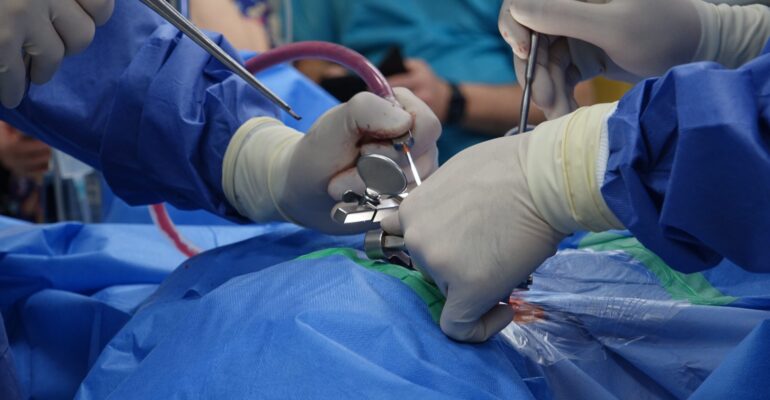
Advanced Pedicle Screw Stabilization Systems
The pedicle screw and rod construct is the dominant spinal stabilization system used worldwide. Pedicle screws achieve fixation by engaging all three columns of the spine simultaneously — providing the most mechanically robust fixation available and enabling the greatest deformity correction forces. The instruments for pedicle screw stabilization systems include:
Advanced Pedicle Screw Entry Tools
The entry into the pedicle is the highest-risk step of posterior spinal stabilization. Advanced instruments for pedicle entry include:
- T-handle awl with depth stop: Allows the surgeon to feel cortical breakthrough at the pedicle isthmus while a depth stop prevents inadvertent over-penetration into the anterior vertebral body cortex
- Curved pedicle finder with tactile feedback shaft: The thin, flexible shaft transmits pedicle wall resistance directly to the surgeon’s hand — an instrument that must be manufactured with consistent rigidity to provide reliable tactile feedback across all cases
- 4-wall ball-tip probe: After pedicle preparation, this instrument systematically checks all four walls (medial, lateral, superior, inferior) and the anterior floor to confirm no breach before screw insertion — the most important safety check in pedicle screw surgery
- Pedicle tap with calibrated markings: Graduated markings along the shaft allow the surgeon to monitor tap depth during cortical bone threading, preventing inadvertent anterior cortex penetration
Advanced Rod System Instruments
The connecting rod is the spine of the fixation construct — it must be contoured precisely to the patient’s sagittal and coronal alignment while maintaining the mechanical strength to resist fatigue failure over decades of use. Advanced rod handling instruments include:
- French bender (in-situ rod bender): Allows final rod adjustment after the rod is partially seated — essential for fine-tuning sagittal alignment once the rod is in place
- Template rod benders: Allow the surgeon to pre-bend the rod to match the patient’s anatomical curvature using a flexible template taken from the patient’s contour — reducing the number of in-situ adjustments needed
- Powerful rod reduction forceps: High-mechanical-advantage instruments that draw the screw head up to the rod level — capable of generating the substantial force required for deformity reduction while maintaining precise control
- Multi-level reduction system: Sequential, multi-segment reduction instruments for complex scoliosis and kyphosis correction — allowing controlled, level-by-level deformity correction across long fusion constructs
- Dual-arm rod holder: Holds the rod in position during reduction without allowing it to rotate or translate — preventing the rod from losing its contour during the reduction maneuver
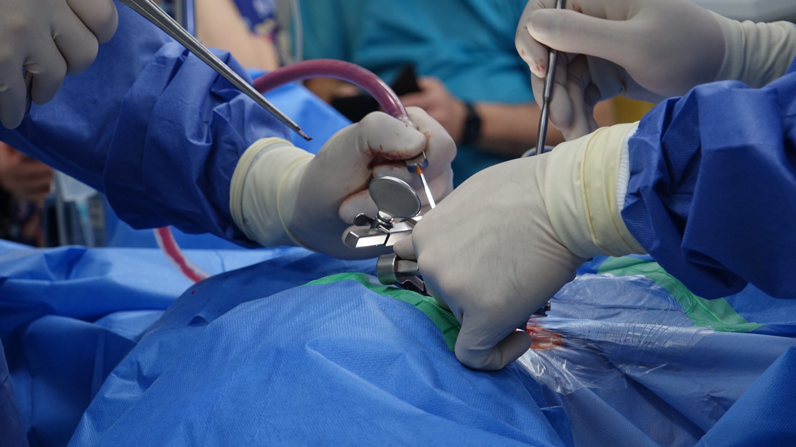
Advanced Decompression Tools for Spine Stabilization Surgery
Safe, effective neural decompression is a prerequisite for successful spinal stabilization. Compression of the spinal cord or nerve roots that remains after stabilization leads to persistent neurological symptoms and poor surgical outcomes. Advanced decompression instruments for stabilization surgery include:
High-Performance Kerrison Rongeurs
The Kerrison rongeur is the primary decompression instrument in spinal stabilization surgery. Key performance specifications for a professional-grade Kerrison rongeur include:
- Carbide jaw inserts: Tungsten carbide cutting inserts maintain sharp cutting edges 3–5× longer than standard stainless steel jaws — critical in high-volume spine programs where rongeurs are used in multiple procedures per day
- Foot plate thickness: The foot plate must be thin enough to slide under bone without tearing the dura, but thick enough to resist breakage during forceful bone removal — a precise manufacturing balance
- Consistent spring tension: The spring mechanism must reliably open the jaw after each bite without requiring the surgeon to manually open it — hand fatigue in long decompression procedures is directly linked to spring quality
- Bite sizes 1–5 mm, angles 40° and 90°: A complete rongeur set must include this full range to address all decompression requirements, from tight lateral recesses (1–2 mm, 90°) to broad central canal decompression (4–5 mm, 40°)
Advanced Disc Removal Instruments
Discectomy — removal of the intervertebral disc — is required for most interbody fusion procedures. Advanced disc removal instruments include:
- Angled disc rongeurs (upbiting, 45° downbiting, straight): The complete set of angled pituitary rongeurs covers all quadrants of the disc space, removing nucleus pulposus material from the anterior, posterior, and lateral disc space without repositioning the retractors
- Long-handled disc rongeurs: Extended-length instruments for L5/S1 and other deep disc spaces where standard-length handles cannot reach the posterior disc through the surgical approach
- Curettes (sizes 1–5, angled 0°, 45°, 90°): For removing cartilaginous end plate, disc material, and granulation tissue from the interbody space — essential for preparing the fusion bed
- Disc space spreaders: Serrated jaw distraction instruments that open collapsed disc spaces before curette and cage insertion — restoring disc height and improving working space
Advanced Nerve Root Protection Instruments
Protecting the nerve root and dura during decompression and interbody cage insertion is one of the highest-priority safety tasks in spine stabilization surgery. Advanced nerve protection instruments include:
- Nerve root retractors (angled and malleable): Fine, flexible instruments that gently mobilize nerve roots and maintain them in a protected position — must have a smooth, atraumatic surface to prevent axonal injury during prolonged retraction
- Penfield dissectors (sizes 1–5): Versatile, blunt-tipped dissectors for mobilizing nerve roots, dissecting epidural adhesions, and creating working space between neural elements and bone — an indispensable set in any spine stabilization procedure
- Dural retractors: Extremely fine, flexible retractors for carefully mobilizing the dural sac during central decompression — must be atraumatic, as dural tears are the most common intraoperative complication in spine decompression surgery
Advanced Interbody Fusion Stabilization Instruments
Interbody fusion — achieving bone growth between adjacent vertebral bodies through a cage placed in the disc space — provides the anterior column support essential for durable spinal stabilization. Advanced interbody fusion instruments include:
End Plate Preparation Instruments
- Powered end plate shavers: Motorized instruments with interchangeable cutting heads that prepare the vertebral end plate to a consistent surface, removing cartilage and creating optimal bleeding bone for fusion — significantly faster and more consistent than manual curettes for high-volume programs
- End plate curettes (angled/straight in graduated sizes): Manual instruments for meticulous end plate preparation when precise tactile feedback is needed — particularly valuable in fragile osteoporotic end plates where over-aggressive preparation risks end plate violation and cage subsidence
- Rasps and files: For final smoothing of the prepared end plate surface before cage insertion — creating the flat, even surface that maximises the cage-bone contact area for fusion
Cage Insertion and Positioning Instruments
- Trial sizers (in full size range): A complete set of trial implants matching every available cage size — allowing systematic footprint sizing to identify the cage that maximises end plate coverage without overhanging the ring apophysis. Trials must be durable enough to withstand repeated impaction
- Cage inserter with anti-rotation locking mechanism: Securely locks to the cage to prevent inadvertent cage rotation during impaction — a critical safety feature that prevents the cage from turning in the disc space and impinging on neural structures
- Angled inserter for TLIF approach: The transforaminal approach requires a specific angled inserter geometry that allows cage delivery through the unilateral posterior approach — a different instrument than the straight PLIF inserter
- Cage impactor with depth stop: Delivers controlled impact force to advance the cage to the correct depth without the risk of over-penetration into the contralateral disc space or anterior longitudinal ligament
Minimally Invasive Spine Stabilization Tools
Minimally invasive spine surgery (MIS) has transformed the field of spinal stabilization — achieving equivalent fixation to open surgery through dramatically smaller incisions, with less blood loss, reduced muscle damage, shorter hospital stays, and faster return to function. MIS stabilization requires a completely different instrument philosophy:
Percutaneous Pedicle Screw System Instruments
- Percutaneous K-wire and guide pin systems: Fluoroscopy-guided guidewires establish the pedicle trajectory before serial dilation — the starting point for all percutaneous pedicle screw systems
- Cannulated dilating trocar sets: Sequential dilators expand the muscle corridor from the K-wire to the diameter needed for percutaneous screw insertion — minimising muscle denervation and devascularization
- Extended-tab screwdrivers: Long, slender screwdrivers that extend 7–10 cm above the skin surface, transmitting torque to the pedicle screw through a minimally invasive working channel without requiring open muscle retraction
- Rod delivery and connection systems (MIS): Specialized instruments for passing the connecting rod through subcutaneous tissue between percutaneous screw tulips — one of the most technically demanding steps in MIS fixation
Tubular Retractor Systems
- Sequential dilating tube sets: Progressively larger dilating tubes expand the paraspinal muscle corridor to the spine via sequential blunt dilation, rather than traditional cutting and retraction
- Working channel retractor tubes: The final tube — typically 18 mm to 26 mm diameter — provides a fixed working channel through which all surgical instruments, decompression tools, and interbody fusion instruments are passed
- Table-mounted retractor arms: Rigid mechanical arms that hold the working channel tube rigidly in position, freeing the surgical assistant from manual tube holding and providing a stable working environment throughout the procedure
- MIS-specific instruments: Extended-length, narrow-profile versions of standard instruments (curettes, rongeurs, nerve retractors) designed to work through the limited diameter of the tubular retractor working channel
Anterior Cervical Stabilization Tools
The cervical spine presents unique stabilization challenges — the confined surgical corridor, proximity of the carotid arteries, vertebral arteries, esophagus, trachea, and the spinal cord at its most vulnerable segment demand instruments of exceptional precision and safety. Professional anterior cervical stabilization tools include:
- Caspar vertebral body distraction pin system: Parallel pins inserted into adjacent vertebral bodies at calibrated spacing, used with a dedicated distractor to open the cervical disc space under controlled, measurable force. The most widely used cervical disc space distraction system in the world
- Self-retaining cervical retractor systems: Handheld and table-mounted retractors that maintain stable retraction of the carotid sheath, esophagus, and trachea throughout the procedure without requiring continuous manual retraction
- Micro Kerrison rongeurs (1–3 mm, upbiting): Specialized small-bite rongeurs for cervical disc fragment removal and foraminal decompression — the narrow cervical canal demands instruments with exceptional precision and minimal footprint
- High-speed micro-drill system: Fine-tipped burrs for removing osteophytes, uncinate spurs, and posterior vertebral body rim without the dural risk of rongeur use in the narrow cervical canal
- Cervical plate holding systems: Instruments that maintain the anterior cervical plate in perfect position during screw insertion — with one hand holding the plate, the other hand must be free to insert and tighten each screw without plate migration
Comparison: Open vs. Minimally Invasive Spine Stabilization Instruments
| Feature | Open Spinal Stabilization | Minimally Invasive (MIS) |
|---|---|---|
| Muscle retraction | Taylor or Gelpi retractors — static retraction | Sequential dilating tubular retractor system |
| Pedicle screw insertion | Standard screwdrivers under direct vision | Percutaneous screwdrivers with extended tabs under fluoroscopy |
| Rod delivery | Direct placement under open vision | Subcutaneous rod delivery through percutaneous connectors |
| Decompression instruments | Standard-length rongeurs, curettes, drills | Extended-length, narrow-profile instruments through tube |
| Blood loss | Higher — muscle stripping and retraction | Significantly lower — muscle dilating approach |
| Recovery time | Longer — 3–5 days hospital; 6–12 weeks recovery | Shorter — 1–2 days hospital; 2–6 weeks recovery |
| Fluoroscopy requirement | Intermittent confirmation imaging | Continuous fluoroscopy or navigation guidance |
| Deformity correction | Excellent — direct manipulation possible | Limited — indirect reduction via screw-rod system only |
| Instrument complexity | Moderate — familiar standard instruments | High — system-specific, technique-dependent instruments |
Patient Safety Features in Advanced Spine Stabilization Tools
The most advanced professional spine stabilization tools incorporate specific safety engineering features that reduce the risk of serious intraoperative complications:
Depth Control Systems
Depth stops on pedicle awls, taps, and screwdrivers prevent instruments from advancing beyond the safe working depth into the vertebral body — reducing the risk of anterior cortex penetration and great vessel injury. Calibrated depth markings on instruments allow the surgeon to monitor depth continuously during instrument advancement.
Anti-Rotation Locking
Cage inserters and screw-holding sleeves with anti-rotation locking mechanisms prevent the implant from rotating during impaction — a critical safety feature since rotation of a cage or screw in the disc space or pedicle can result in nerve root impingement or dural tear.
Tactile Feedback Optimization
Professional pedicle probes and awls are designed with specific shaft flexibility and stiffness characteristics that optimize the transmission of tactile feedback to the surgeon’s hand. The surgeon’s ability to feel the difference between pedicle cancellous bone and cortical wall is the primary safety mechanism in pedicle screw placement — and it depends entirely on the mechanical properties of the probe.
Torque Limitation
Torque-limiting screwdrivers in spine stabilization systems prevent pedicle screw overtightening — a significant risk in osteoporotic bone where excessive insertion torque strips the pedicle and converts a stable fixation into an unstable one. Pre-set torque limiters provide an audible click when the maximum safe torque is reached.
Instrument Counting and Tracking
Organised instrument trays with clearly labeled, individual instrument slots support rigorous surgical count management. In spine surgery — where small instruments operate near the spinal cord — a retained instrument can be catastrophic. Professional spine stabilization instrument sets use colour-coded trays, numbered slots, and silhouette marking to enable immediate visual identification of any missing instrument.
Quality Standards for Professional Spine Stabilization Tools
| Quality Criterion | Professional Standard | Consequence of Non-Compliance |
|---|---|---|
| Material grade | 316L surgical stainless steel — certified | Corrosion, dimensional instability, ion release |
| Dimensional tolerance | ±0.05 mm for critical measurement instruments | Incorrect screw length selection, end plate violation |
| Cutting edge hardness | Carbide inserts for rongeurs and curettes | Rapid dulling, incomplete decompression |
| Surface finish | Mirror or satin — no rough surfaces | Biofilm harboring, inadequate sterilization |
| Autoclave cycles | 500+ at 134°C without performance loss | Instrument failure during procedure |
| Regulatory compliance | ISO 13485, CE marking, FDA clearance | Legal liability, quality management gaps |
| Implant compatibility | Matched to specific screw/rod system | Cam-out, stripped screws, inadequate fixation |
| Tactile feedback | Validated shaft rigidity and tip sensitivity | Pedicle wall breach, neurological injury |
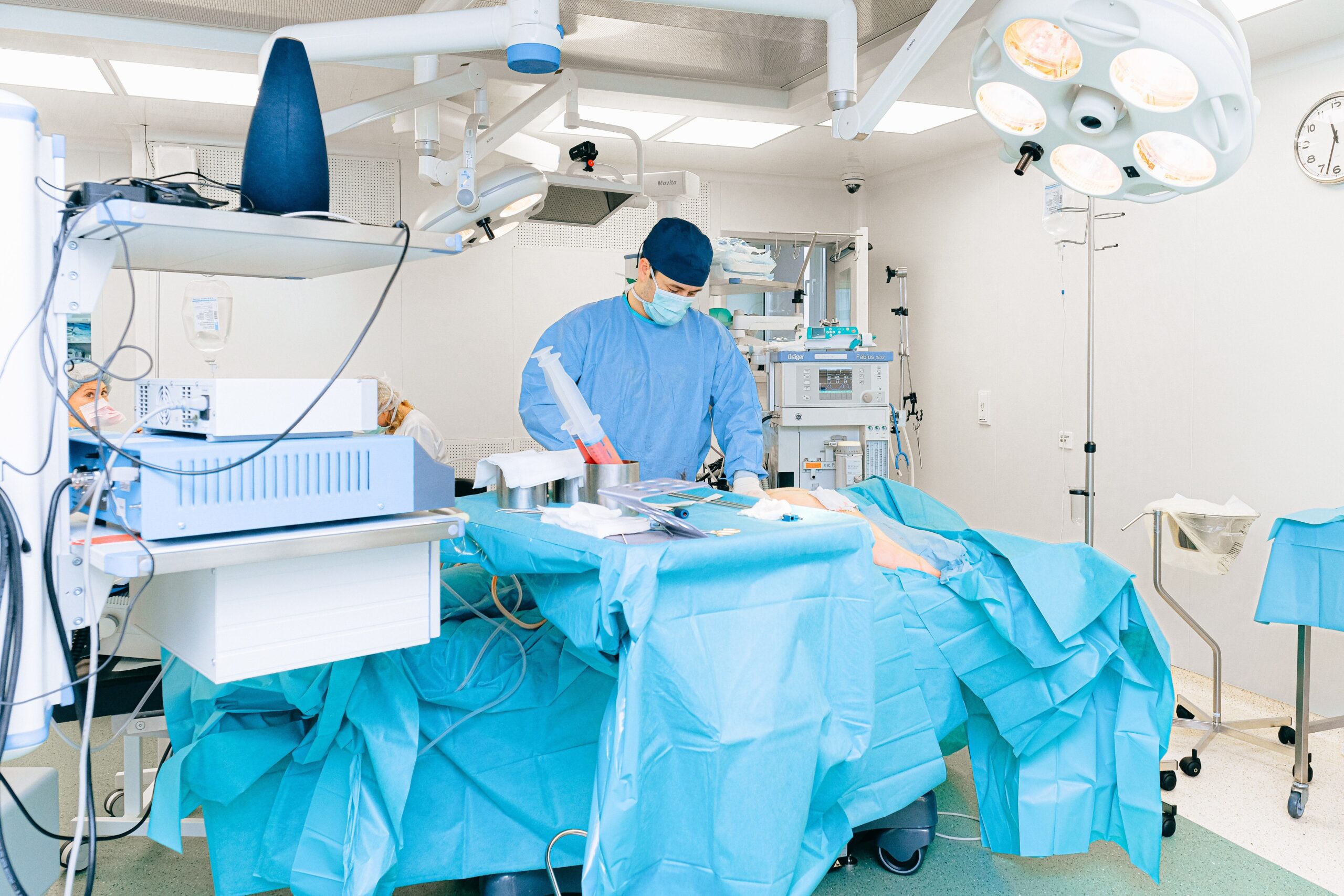
Setting Up a Spine Stabilization Instrument Program: Key Considerations
For hospitals and surgical centers establishing or upgrading a spine stabilization program, the instrument acquisition strategy is as important as the implant system selection. Key considerations include:
Instrument-Implant System Matching
Every pedicle screw system requires instruments matched to its specific screw drive geometry, rod diameter, and tulip dimensions. Mismatched instruments are not just inefficient — they are dangerous. A screwdriver that doesn’t precisely fit the screw drive will cam out, stripping the screw head and potentially leaving a screw that cannot be advanced or removed. Before purchasing instruments, confirm exact compatibility with the implant systems used in your program.
Instrument Set Configuration
Configure instrument sets for each procedure type — a thoracolumbar fusion set, a cervical fusion set, a microdiscectomy set, and a deformity correction set may all be required. Avoid the temptation to use a single set for all procedures — instrument configurations that are “close enough” for one procedure may be genuinely inadequate for another.
Backup Sets
High-volume spine programs should maintain at least two complete sets for each procedure type — one in use and one being sterilized. Single-set programs create bottlenecks in high-volume environments and leave the program exposed to unacceptable delays if an instrument is damaged or contaminated during a case.
Instrument Maintenance Program
Establish a formal instrument inspection program — every instrument checked before each procedure, Kerrison rongeurs sharpness tested quarterly, probe tips inspected for bending, and screwdriver tips checked for cam-out risk. Replace worn instruments before they fail — the cost of a replaced instrument is always less than the cost of an intraoperative complication.
Why Hasni Surgical for Professional Spine Stabilization Tools?
Hasni Surgical manufactures and supplies professional spine stabilization instruments to spine surgery programs, hospitals, and surgical centers worldwide. Our spine instrument range is built on the same uncompromising commitment to precision, material quality, and clinical reliability that defines every instrument we produce.
- ✅ 316L surgical stainless steel — certified: Every spine instrument manufactured from documented, certified surgical-grade steel
- ✅ ISO 13485 certified manufacturing: Quality management system audited and certified to international medical device standards
- ✅ Complete spine surgery sets: 28–35 piece comprehensive spinal surgery sets with autoclavable sterilizing boxes for organized deployment
- ✅ Individual instrument supply: Any instrument in our spine range available individually for set completion or single-item replacement
- ✅ Brand new — every unit: No refurbished or reprocessed instruments ever supplied
- ✅ Ultrasonic cleaned before packaging: Instruments arrive clean and ready for final sterilization without additional decontamination
- ✅ CE and ISO certified products: Spine instrument sets certified to international medical device standards
- ✅ Worldwide shipping — dispatched within 2 working days: Fast delivery to hospitals and clinics in more than 50 countries
- ✅ 25% bulk order discount: Significant savings for hospitals, surgical centers, and distributors purchasing in volume
- ✅ 60-day money-back guarantee: Complete confidence in quality — full refund if not satisfied
Frequently Asked Questions (FAQ)
What are spine stabilization tools?
Spine stabilization tools are specialized surgical instruments used to achieve mechanical stabilization of the vertebral column — through pedicle screw and rod fixation, interbody cage placement, anterior plate fixation, or a combination of these. They include pedicle awls, finders, probes, taps, screwdrivers, rod benders and reducers, Kerrison rongeurs, disc curettes, nerve retractors, cage inserters, and complete system instruments — all working together to achieve safe, precise spinal stabilization while protecting neural structures.
What is the difference between spinal fixation and spinal fusion?
Spinal fixation refers to the mechanical immobilization of vertebral segments using metallic implants (pedicle screws, rods, plates, cages) — it provides immediate stability. Spinal fusion refers to the biological process of bone growing across the target motion segment, creating a permanent rigid bridge between vertebrae — this takes 3–12 months to achieve after surgery. Fixation supports the spine while fusion occurs; fusion provides the permanent biological stability that allows fixation implants to eventually be unloaded.
What makes a pedicle probe the most important safety instrument in spine surgery?
The pedicle probe (ball-tip probe) is used after pedicle preparation to tactilely verify that all four cortical walls of the pedicle remain intact before a pedicle screw is inserted. A pedicle breach — particularly medial wall breach — can place a screw into the spinal canal, causing direct cord or nerve root injury. The probe’s ability to detect this before screw insertion prevents one of the most serious complications in spinal fixation surgery. Its safety value makes it the single most important non-cutting instrument in the spine surgical set.
Why are carbide jaw inserts important in Kerrison rongeurs for spine surgery?
Tungsten carbide cutting inserts in Kerrison rongeur jaws maintain sharp cutting edges 3–5 times longer than standard stainless steel jaws. A dull rongeur requires more force per bite — increasing the risk of slipping, dural laceration, and nerve root injury. In high-volume spine programs, carbide-insert rongeurs dramatically reduce the frequency of blade sharpening or replacement, lowering total instrument cost while maintaining consistent cutting performance and patient safety.
Can spine stabilization instruments be used for both cervical and lumbar surgery?
Some instruments are shared between cervical and lumbar spine surgery — Cobb elevators, Penfield dissectors, basic curettes, and certain retractors. However, many instruments are anatomy-specific: cervical disc rongeurs are smaller and angled differently than lumbar rongeurs; cervical screw systems use different drive geometries than thoracolumbar pedicle screw systems; and cage inserters are approach and size specific. Most spine programs maintain separate cervical and thoracolumbar instrument sets to ensure the correct instruments are available for each procedure.
How should spine stabilization instruments be organized for OR efficiency?
Professional spine stabilization instruments should be stored in labeled, individually slotted cassette trays within autoclavable sterilizing boxes. Trays should be organized by surgical workflow — pedicle access instruments together, rod system instruments together, decompression instruments together — so that scrub technicians can hand instruments to the surgeon in logical sequence without searching. Color-coding by instrument category and numbered slots that match instrument count sheets enable rapid pre- and post-procedure counting, reducing the risk of retained instruments.
Conclusion
Professional spine stabilization tools represent the highest standard of surgical instrument engineering — demanded by the most unforgiving surgical environment in orthopedic surgery. When instruments are used within centimeters of the spinal cord, in a deep surgical field under time pressure, with a patient’s neurological function and quality of life at stake — there is simply no room for instrument imprecision, mechanical failure, or quality compromise.
From advanced pedicle access tools with optimized tactile feedback, to high-performance carbide-insert Kerrison rongeurs for safe decompression, to sophisticated rod reduction systems for complex deformity correction, to the emerging world of MIS stabilization instruments — every tool in the spine stabilization set exists to make the surgery safer for the patient and the surgeon.
Explore Hasni Surgical’s complete range of professional spine stabilization tools — certified quality, worldwide shipping, and the manufacturing expertise of Sialkot’s world-class surgical instrument industry, dedicated to every spine surgery program we serve.