
What Is an Otoscope? Complete Guide to Types, Uses & Buying the Best One
June 23, 2026 2026-06-23 4:23What Is an Otoscope? Complete Guide to Types, Uses & Buying the Best One
What Is an Otoscope? Complete Guide to Types, Uses & Buying the Best One

Have you ever sat in a doctor’s office with an earache, and watched them pick up a small handheld instrument, shine a light into your ear, and within seconds tell you exactly what was wrong? That instrument is an otoscope — and it is one of the most important diagnostic tools in all of modern medicine.
Used every single day by ENT specialists, general practitioners, pediatricians, audiologists, emergency physicians, nurses, and veterinarians, the otoscope provides a direct, magnified, illuminated view of the ear canal and eardrum — a view that is simply not possible any other way. Despite how critical this instrument is, many clinicians, medical students, and buyers know surprisingly little about how otoscopes work, what types are available, and how to choose the right one.
This comprehensive guide covers everything: what an otoscope is, how it works, the six major types, which conditions it diagnoses, correct examination technique, LED versus halogen comparison, maintenance tips, and a complete buyer’s checklist.
“The otoscope is to the ear what the stethoscope is to the heart — an irreplaceable clinical window into a part of the body that cannot otherwise be seen.”
What Is an Otoscope?
An otoscope — also called an auriscope — is a handheld medical diagnostic instrument designed to examine the external ear canal and the tympanic membrane (eardrum). It combines a light source, a magnifying lens, and a funnel-shaped speculum tip to allow clinicians to visualize the interior of the ear that would otherwise be completely inaccessible.
The name comes from the Greek words oto (ear) and skopein (to examine). First developed in the early 19th century, the otoscope has evolved from a simple candle-and-mirror arrangement into sophisticated digital instruments with HD cameras, wireless connectivity, and real-time image sharing.
In a single 30-second examination, a trained clinician using an otoscope can identify:
- Ear infections — both otitis media (middle ear) and otitis externa (outer ear canal)
- Eardrum perforations, scarring, or retraction
- Fluid behind the eardrum (otitis media with effusion / glue ear)
- Earwax (cerumen) impaction blocking the ear canal
- Foreign bodies lodged in the ear canal
- Cholesteatoma — an abnormal destructive skin growth behind the eardrum
- Ear canal tumors or polyps
- Signs of eustachian tube dysfunction
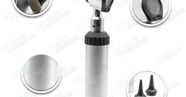
Anatomy of an Otoscope: Key Components

1. Handle
The handle is the body of the otoscope, housing the power source — either standard AA or C batteries, or a rechargeable lithium-ion battery system. Premium handles feature textured or rubberized grips to prevent slipping and are ergonomically contoured for comfortable one-handed use during extended examination sessions.
2. Head
The head of the otoscope contains the light source and magnifying lens system. It attaches to the handle and is the most technically precise component of the instrument. Standard heads provide 3× magnification; specialist ENT models offer up to 5× for fine-detail tympanic membrane assessment.
3. Light Source
The quality of illumination directly determines examination quality. Modern professional otoscopes use LED or fiber-optic light sources that produce bright, white, daylight-quality illumination — rendering tissue colors accurately so that subtle findings like early eardrum redness, amber-colored fluid, or pale retraction are clearly visible. Older halogen bulb otoscopes produce a yellowish light that can mask early pathology.
4. Magnifying Lens
A removable magnifying lens sits at the viewing end of the head. Standard models provide 3× magnification — sufficient for most clinical needs. Some models include a pneumatic port adjacent to the lens that allows a small puff of air to be directed into the sealed ear canal to assess eardrum mobility — an essential feature for diagnosing middle ear fluid in children.
5. Ear Speculum
The speculum is the funnel-shaped tip that attaches to the front of the otoscope head. It gently enters the outer portion of the ear canal to direct light and focus the field of view. Specula come in multiple sizes — 2.5 mm, 3 mm, 4 mm, and 5 mm — to accommodate different ear canal sizes from newborns to adults. Most professional kits include all sizes.
6 Types of Otoscopes: Which One Do You Need?

Type 1: Standard (Conventional) Otoscope
The most widely used otoscope type in primary care, pediatrics, emergency medicine, and general practice globally. Standard otoscopes provide a direct view through the magnifying lens and are available in both battery-powered and rechargeable versions. Reliable, affordable, and effective in trained hands — the foundation of ear examination worldwide.
- ✅ Best for: General practice, primary care, emergency departments, pediatrics, nursing
- ✅ Advantages: Cost-effective, reliable, widely available, easy to learn
- ⚠️ Limitation: Only the examiner sees the image; no digital recording capability
Type 2: Pneumatic Otoscope
The pneumatic otoscope adds a rubber bulb and connecting tube that allows the clinician to puff a small amount of air into the sealed ear canal during examination. This tests tympanic membrane mobility — a freely moving eardrum indicates a healthy middle ear, while reduced movement suggests the presence of fluid (otitis media with effusion) or other middle ear pathology. Essential equipment for any pediatrician.
- ✅ Best for: Pediatricians, OME diagnosis, assessing eardrum mobility
- ✅ Advantages: Adds functional assessment to visual examination; essential for glue ear diagnosis
- ⚠️ Limitation: Requires airtight speculum seal; results are technique-dependent

Type 3: Digital / Video Otoscope
Digital otoscopes incorporate a miniature camera in the head that transmits a live, high-definition image to a connected screen — a built-in display, smartphone, tablet, or computer via USB or WiFi. This technology allows both the clinician and the patient to see the ear simultaneously, dramatically improving patient education and communication. Images and videos can be saved for medical records, shared with distant specialists, or used in telemedicine consultations.
- ✅ Best for: Telemedicine, patient education, documentation, specialist referrals, home use
- ✅ Advantages: Shareable HD images; patient sees their own ear in real time; excellent for training
- ⚠️ Limitation: Higher cost; requires charged battery and connectivity setup
Type 4: Fiber-Optic Otoscope
Fiber-optic otoscopes transmit light via bundles of optical glass fibers from the handle to the examination field — rather than having the bulb in the head itself. This delivers extremely bright, shadow-free, cool, white illumination directly to the ear canal. The superior light quality renders tissue colors with exceptional accuracy, making fiber-optic otoscopes the preferred choice of ENT specialists who require the finest diagnostic detail.
- ✅ Best for: ENT specialists, high-volume clinical practice, fine tympanic membrane assessment
- ✅ Advantages: Superior light quality; cooler head; best-in-class tissue color rendering
- ⚠️ Limitation: Higher cost; fiber bundles can degrade if kinked or mishandled

Type 5: Rigid Endoscopic Otoscope
Rigid endoscopic otoscopes use a narrow rod-lens endoscope — typically 2.7 mm or 4 mm in diameter — connected to a dedicated HD camera and external light source. They provide the highest quality visualization of the ear canal and middle ear of any otoscope type, producing near-surgical-microscope clarity when displayed on a high-definition monitor. Available in 0°, 30°, and 70° angled configurations for different viewing requirements.
- ✅ Best for: ENT surgeons, operating room use, endoscopic ear surgery, advanced diagnostics
- ✅ Advantages: Highest image quality; angled views of hidden anatomy; surgical-grade visualization
- ⚠️ Limitation: Expensive; requires external light source and camera system; specialist use only
Type 6: Veterinary Otoscope
Veterinary otoscopes are purpose-built for examining the ears of animals. Animal ear canals differ substantially from human ears — they are often longer, more acutely angled (particularly in dogs), and vary enormously in size across species. Veterinary otoscopes include longer, wider specula, brighter light sources, and more durable construction for the demands of animal examination. They are essential for diagnosing otitis, ear mites, foreign bodies, polyps, and canal tumors in dogs, cats, horses, rabbits, and exotic species.
- ✅ Best for: Veterinary clinics, animal hospitals, mobile vets, exotic animal practices
- ✅ Advantages: Longer specula for deep access; robust for animal handling; bright illumination for thickly haired canals
- ⚠️ Limitation: Not suitable for human use due to speculum size differences
Common Conditions Diagnosed with an Otoscope
Acute Otitis Media (AOM) — Middle Ear Infection
The most common reason for otoscopy in children. AOM is a bacterial or viral infection of the middle ear space behind the eardrum. On otoscopy, the eardrum appears red, bulging, and opaque rather than its normal translucent grey-white appearance — and there may be visible pus or yellow discharge. AOM is the number one reason for antibiotic prescriptions in children worldwide, and accurate otoscopic diagnosis is essential to avoid unnecessary antibiotic use.
Otitis Media with Effusion (OME) — Glue Ear
Non-infectious fluid accumulation in the middle ear, common after AOM or due to eustachian tube dysfunction. The eardrum may appear amber or yellowish, retracted, or show visible air-fluid levels or bubbles behind it. Pneumatic otoscopy confirms diagnosis — reduced eardrum mobility is the key finding. OME is a leading cause of temporary hearing loss and speech delay in children under 5.
Otitis Externa — Swimmer’s Ear
A bacterial infection of the outer ear canal itself, most commonly caused by Pseudomonas aeruginosa following water exposure. On otoscopy the canal walls appear red, swollen, and covered with debris or discharge — the canal may be so edematous that the eardrum cannot even be visualized. Otoscopy is critical for distinguishing otitis externa from otitis media, as these conditions require completely different treatments.
Cerumen (Earwax) Impaction
Accumulated earwax blocking the ear canal is one of the most common findings on routine otoscopy — affecting approximately 6% of the general population. It causes hearing loss, tinnitus, ear pain, dizziness, and a sensation of fullness. Otoscopy immediately confirms whether the canal is clear or obstructed, and guides the appropriate removal method — irrigation, microsuction, or softening drops.
Tympanic Membrane Perforation
A hole in the eardrum from infection, blunt trauma, blast injury, barotrauma, or previous surgery. Otoscopy reveals a dark defect in the otherwise translucent eardrum. The size and position of the perforation guide treatment — central perforations often heal spontaneously, while marginal perforations or those associated with cholesteatoma require surgical repair.
Cholesteatoma
An aggressive, destructive growth of keratinizing squamous epithelium within the middle ear or mastoid — often arising from a retraction pocket in the pars flaccida of the eardrum. Without surgical treatment, cholesteatoma erodes bone including the ossicles, facial nerve canal, semicircular canals, and skull base. Early detection on careful otoscopic examination is lifesaving — a small retraction pocket or attic crust on otoscopy should never be dismissed.
Foreign Bodies
Particularly common in young children who insert objects — beads, erasers, cotton wool, insects, seeds, and food — into their ear canals. Otoscopy identifies the foreign body, its exact location, and its proximity to the eardrum — all critical information for selecting the safest removal technique and avoiding iatrogenic eardrum injury.
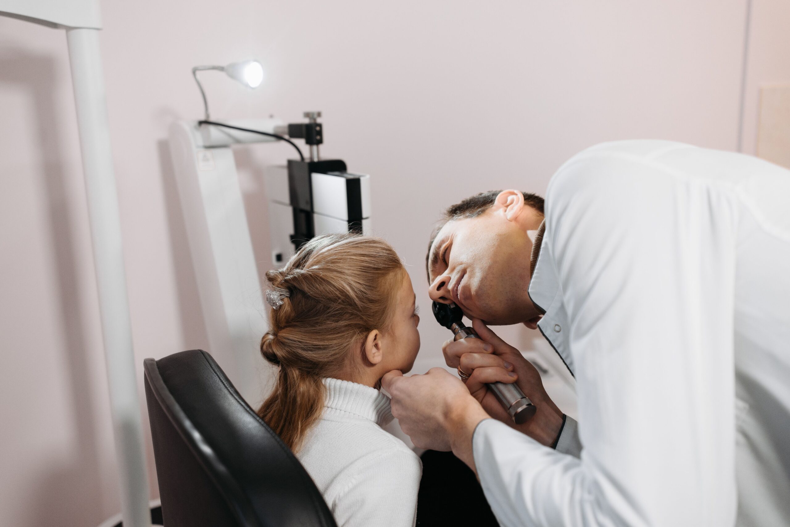
How to Use an Otoscope Correctly: Step-by-Step

Correct otoscopic technique is a core clinical skill. Improper use leads to patient discomfort, missed pathology, and potentially false-reassurance in serious conditions. Follow this evidence-based technique:
- Select the correct speculum size: Choose the largest speculum that fits comfortably in the patient’s ear canal — typically 4–5 mm for adults, 3 mm for older children, 2.5 mm for infants. Attach it firmly to the otoscope head.
- Check your light source: Turn on and confirm the light is bright and consistent before inserting into the ear. Never begin an examination with a dim or flickering light — it is the leading cause of missed otoscopic diagnoses.
- Straighten the ear canal: In adults and older children, gently pull the pinna (outer ear) upward and backward to straighten the naturally curved ear canal. In children under age 3, pull the pinna gently downward and backward.
- Insert the speculum carefully: Insert the speculum into the outer portion of the ear canal at a slight downward and forward angle, following the natural anatomy. Never force insertion — the speculum should slip in smoothly and painlessly.
- Stabilize your hand against the patient’s head: Rest the hand holding the otoscope gently against the patient’s cheek or temporal region. This is a non-negotiable safety measure — if the patient moves unexpectedly, your hand moves with them, preventing the speculum from being inadvertently driven deeper.
- Systematically examine the canal then eardrum: First inspect the ear canal walls for redness, swelling, discharge, or foreign bodies. Then advance focus to the eardrum and identify key landmarks: the light reflex (cone of light), the handle and short process of the malleus, the pars tensa, and the pars flaccida.
- Pneumatic assessment if indicated: For suspected otitis media with effusion, attach the pneumatic bulb, create a gentle airtight seal with the speculum, and assess eardrum mobility by giving a gentle squeeze and release of the bulb.
- Document all findings: Record side examined, appearance of ear canal and eardrum, specific landmarks identified, and any abnormalities noted. Good documentation protects the clinician and supports continuity of care.
LED vs. Halogen Otoscopes: A Detailed Comparison

| Feature | LED Otoscope | Halogen Otoscope |
|---|---|---|
| Light color temperature | 5000–6500K — bright white / daylight quality | 3000–3500K — warm yellow-white |
| Tissue color accuracy | Excellent — redness, amber fluid, and grey tones rendered accurately | Good but yellow cast can mask early eardrum redness |
| Bulb lifespan | 50,000+ hours — virtually maintenance-free | Hundreds of hours — bulbs need regular replacement |
| Heat generated | Minimal — LED runs cool throughout examination | More heat — can cause minor discomfort with prolonged speculum dwell |
| Brightness output | Higher lumens — better penetration in wax-filled or narrow canals | Adequate for most examinations but dimmer |
| Battery efficiency | Much lower power draw — longer battery life per charge | Higher power draw — batteries drain faster |
| Purchase cost | Slightly higher initial cost | Lower initial cost |
| Running cost | Very low — no bulb replacement needed | Ongoing bulb replacement adds up over time |
| Recommendation | ✅ Strongly recommended for all new purchases | Acceptable only if budget constraints prevent LED purchase |
The clinical case for LED over halogen is compelling. The superior white light of LED otoscopes makes it significantly easier to detect subtle early pathology — particularly the slight pink blush of early AOM, the amber tinge of middle ear fluid, and the pale retracted appearance of eustachian tube dysfunction. For any clinician purchasing a new otoscope in 2024 or beyond, LED is the clear and unambiguous choice.
Otoscope & Ophthalmoscope Combination Diagnostic Sets

Many clinicians purchase combination diagnostic sets that include both an otoscope and an ophthalmoscope sharing a single rechargeable handle and charging base. These combination sets are the standard toolkit for:
- Medical and nursing students learning systematic clinical examination
- General practitioners who perform routine ear and eye examinations in the same consultation
- Pediatricians who examine ears, eyes, nose, and throat at every well-child check
- Emergency physicians who need rapid head-to-toe assessment capability
- Urgent care centers with diverse daily patient presentations
Combination sets represent excellent value — one charging system for two critical instruments, with matched optical quality and a consistent user experience. Hasni Surgical’s diagnostic combination sets include both instruments with full speculum sets, a rechargeable handle, and a protective case.
Buyer’s Guide: 10 Things to Check Before You Buy an Otoscope
- ☑️ Light source type: Always choose LED — superior brightness, color accuracy, and lifespan over halogen
- ☑️ Magnification level: 3× for general clinical use; 5× for detailed ENT specialist assessment
- ☑️ Power source: Rechargeable lithium-ion for high-volume clinical settings; standard batteries for portability and home use
- ☑️ Speculum sizes included: Confirm the kit includes adult (4–5 mm), pediatric (3 mm), and infant (2.5 mm) sizes
- ☑️ Pneumatic capability: Essential for any pediatrician diagnosing OME in children
- ☑️ Digital/video option: Consider if telemedicine, patient education, or image documentation is important in your practice
- ☑️ Build quality: All-metal head and handle preferred over plastic — more durable and longer-lasting in clinical environments
- ☑️ Ergonomics: Handle should balance well and feel comfortable for extended use without hand fatigue
- ☑️ International certifications: FDA clearance and CE marking confirm compliance with medical device safety standards
- ☑️ Warranty and support: Choose suppliers with documented warranty terms and responsive customer service
Otoscope Care and Maintenance
A professional otoscope is a precision medical instrument and a long-term investment. Follow these maintenance guidelines to protect its performance and extend its service life:
- Clean after every patient: Wipe the body and head with a 70% isopropyl alcohol wipe after each use. Never immerse any part of the otoscope in liquid — moisture in the optical and electrical components causes permanent damage.
- Use disposable specula in clinical settings: Single-use disposable specula eliminate cross-infection risk entirely. If reusable specula are used, they must be sterilized between each patient per facility infection control protocols.
- Clean the viewing lens gently: Use only a soft, lint-free optical cloth. Never use paper towels or tissues — they scratch optical-grade glass and permanently impair visualization quality.
- Check light output before every clinical session: Dim, inconsistent, or flickering illumination indicates a low battery (in rechargeable models) or an aging bulb (in halogen models). Always examine with a fully bright light.
- Store in the provided case: Keep the otoscope in its protective case or on its charging stand when not in use. Avoid dropping or impacts — the precision optical alignment in the head can be disrupted by even moderate knocks.
- Maintain the battery in rechargeable models: Keep the battery regularly topped up. Allowing lithium-ion batteries to fully discharge repeatedly accelerates capacity loss and shortens battery lifespan significantly.
Frequently Asked Questions (FAQ)
What is an otoscope and what is it used for?
An otoscope is a handheld medical instrument that combines a light source, magnifying lens, and ear speculum to allow clinicians to examine the external ear canal and tympanic membrane (eardrum). It is used to diagnose ear infections, earwax impaction, eardrum perforations, middle ear fluid, foreign bodies, cholesteatoma, and other ear canal conditions.
What is the difference between an otoscope and an ophthalmoscope?
An otoscope examines the ear canal and eardrum. An ophthalmoscope examines the interior of the eye — particularly the retina, optic disc, macula, and retinal blood vessels. Although they have similar handheld designs and are often sold as combination sets sharing a handle, they are completely different instruments used on different anatomical regions.
What does a normal eardrum look like through an otoscope?
A normal tympanic membrane appears pearly grey-white, semi-transparent, and slightly concave (pulled inward). You can see the handle of the malleus running diagonally across it, and the cone of light (light reflex) should be visible pointing anteroinferiorly from the umbo. The eardrum surface is smooth, intact, and moves freely with pneumatic assessment.
Is an otoscope safe to use at home?
Yes — consumer-grade digital otoscopes are safe for home monitoring when used correctly. Key safety rules: insert only the very tip of the speculum, never use in an ear with known perforation, do not force insertion if the child is resisting. Home otoscopy is valuable for monitoring between doctor visits but does not replace professional clinical diagnosis — always seek medical advice for any ear symptoms or treatment decisions.
Why is LED better than halogen in an otoscope?
LED produces bright, white, daylight-quality light (5000–6500K) that renders ear tissue colors accurately — making subtle findings like early eardrum redness, amber middle ear fluid, or pale retraction clearly visible. Halogen produces a warmer yellow light that can mask early pathology. LED bulbs also last 50,000+ hours versus hundreds of hours for halogen, eliminating the cost and inconvenience of bulb replacement.
What certifications should a professional otoscope have?
Any otoscope intended for clinical use should carry CE marking (European medical device compliance) and FDA clearance (US medical device approval). ISO 13485 certification of the manufacturer’s quality management system is an additional assurance of consistent manufacturing standards. Never use uncertified instruments for patient examination.
Does Hasni Surgical offer otoscopes with worldwide shipping?
Yes. Hasni Surgical offers a complete range of professional otoscopes — including LED diagnostic sets, digital otoscopes, combination otoscope-ophthalmoscope kits, rigid endoscopic ear diagnostic sets, and veterinary otoscope kits — all available with worldwide shipping from Sialkot, Pakistan. Orders are typically dispatched within 2 working days, and bulk orders qualify for significant discounts.
Conclusion
The otoscope is one of the most powerful and underappreciated diagnostic instruments in clinical medicine. Portable enough to fit in a coat pocket, yet capable of revealing pathology that changes the course of a patient’s treatment — it is the primary tool for every ear examination at every level of healthcare.
Understanding the types, components, correct technique, and buying criteria ensures that clinicians, procurement teams, students, and patients can make informed decisions about this essential instrument. Whether you need a standard LED otoscope for general practice, a pneumatic model for pediatrics, a digital otoscope for telemedicine, a rigid endoscope for the operating room, or a veterinary set for your animal practice — choosing the right instrument from a trusted supplier makes every examination more accurate, more comfortable, and more clinically valuable.
Explore the full range of professional otoscopes and ENT diagnostic instruments at Hasni Surgical — premium quality, worldwide shipping, and competitive pricing backed by years of surgical instrument manufacturing expertise from Sialkot, Pakistan.