Early Detection with Oral Cancer Screening
August 5, 2024 2024-11-21 6:25Early Detection with Oral Cancer Screening
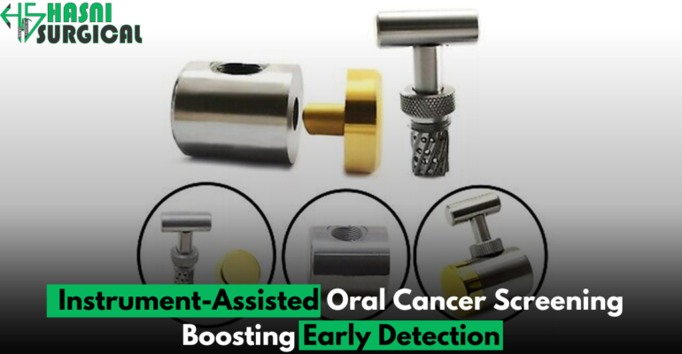
Early Detection with Oral Cancer Screening
Oral Cancer Screening
Cancer, a severe threat to health, often requires early detection and proactive treatment for success. Oral cancer, a major global concern, ranks among the top ten cancers worldwide, with over 657,000 new cases each year. Its prognosis is relatively favorable if caught early, highlighting the need for early detection.
Traditionally, oral cancer screening depended on healthcare professionals carefully inspecting the oral cavity for abnormalities. While this approach has undoubtedly saved lives, its limitations have become increasingly apparent. Subtle lesions or precancerous changes often eluded detection through visual examination alone, necessitating the exploration of supplementary screening methods.
In response, the healthcare community has adopted advanced instrument-assisted screening techniques to improve oral cancer detection. These innovations significantly enhance preventive care, offering clinicians improved diagnostic tools and enabling faster interventions for at–risk individuals.
In this exploration of instrument-assisted oral cancer screening, we examine the evolution, mechanisms, and impact of these methods. From toluidine blue to ViziLite® Plus and VELscope®, we cover key tools for early oral cancer detection.
Explore the future of oral cancer detection with instrument-assisted screening to enhance global health.
Toluidine Blue Staining: Shedding Light on Suspicious Lesions
For early oral cancer detection, healthcare professionals use various tools to identify suspicious lesions. Toluidine blue staining is a key method for enhancing visual examination by highlighting potentially cancerous tissues.
Toluidine blue stains acidic tissue components, highlighting abnormal cells in the oral mucosa. Abnormal areas appear blue or purple, making them easy to spot.
Toluidine blue staining is a quick, routine exam procedure. Here’s how it typically works:
1. Preparation: Before applying Toluidine Blue, examine the oral cavity for any suspicious lesions or concerns. This initial check guides the dye application and ensures a thorough evaluation of problematic areas.
2. Application of Toluidine Blue: Apply a diluted Toluidine Blue solution to the oral mucosa with a swab or brush and leave it for 1-2 minutes. This process stains any abnormal cells present.
3. Visualization: After the allotted time, examine the oral cavity under proper lighting for stain patterns. Abnormalities that retain Toluidine Blue dye appear as dark blue or purple areas against healthier tissue, making them more noticeable.
4. Interpretation: Healthcare providers check Toluidine Blue staining in the oral cavity for persistent dye retention or abnormalities. Suspicious lesions that exhibit strong or irregular staining patterns may warrant further investigation through biopsy or additional diagnostic testing.
Toluidine blue staining offers several advantages as an adjunctive screening tool for oral cancer detection:
Enhanced Visualization: Toluidine Blue stains abnormal cells, highlighting lesions and improving tissue contrast.
Quick and Non-Invasive: The Toluidine Blue staining procedure is quick and easy, needing minimal equipment and no special training. It can be easily incorporated into routine dental or medical check-ups without causing significant discomfort to the patient.
Cost-Effective: Toluidine blue staining is an affordable, efficient diagnostic method that doesn’t need costly equipment.
Complementary to Visual Examination: Toluidine blue staining helps detect lesions not visible during routine exams.
Toluidine blue staining helps detect abnormal cells early but isn’t a standalone diagnostic test. Positive results suggest abnormalities, not cancer. A biopsy and histopathological analysis are needed for a definitive diagnosis and treatment plan.
In conclusion, toluidine blue staining is a valuable tool for early oral cancer detection. It highlights abnormal cells, making suspicious lesions more visible and guiding further diagnostic evaluation, which can improve patient outcomes.
Expanding on ViziLite® Plus: Illuminating Oral Cancer Detection
ViziLite® Plus is a cutting-edge oral cancer screening technology that improves the detection of abnormalities. It uses chemiluminescent light and acetic acid to highlight early signs of oral cancer and precancerous lesions.
ViziLite® Plus uses a chemiluminescent light source that emits a specific wavelength to better visualize oral mucosal abnormalities. This light makes potential lesions or abnormalities fluoresce, contrasting with healthy tissue. This allows healthcare providers to spot and evaluate suspicious lesions more effectively.
The effectiveness of ViziLite® Plus is further amplified by the use of an acetic acid rinse prior to examination. Acetic acid, or vinegar, temporarily changes the appearance of oral mucosal tissues, enhancing visibility of abnormalities. When used with ViziLite®’s chemiluminescent light, it creates a sensitive screening environment to detect early-stage oral cancer or precancerous conditions.
One of the key advantages of ViziLite® Plus is its ease of use and non-invasive nature. The screening process is quick and straightforward, typically taking just a few minutes to complete. Patients rinse their mouths with an acetic acid solution, highlighting abnormalities before examination with the ViziLite® light. This minimally invasive procedure requires no special preparation and can be easily included in routine dental and medical check-ups.
In addition to its effectiveness as a screening tool, ViziLite® Plus offers another crucial benefit: peace of mind. Advanced tools like ViziLite® Plus can boost confidence in oral cancer screening. By detecting abnormalities early, ViziLite® Plus helps both patients and providers take proactive steps for better outcomes.
In conclusion, ViziLite® Plus represents a significant advancement in oral cancer screening by combining chemiluminescent light and acetic acid rinse. Its ease of use, non-invasiveness, and precision improve early diagnosis and outcomes for oral cancer.
The VELscope® system from LED Dental Inc. uses fluorescence to enhance early detection of oral cancer.
The VELscope® system features a handheld device that emits a safe blue light into the oral cavity. This specialized light interacts with the tissues of the mouth, causing certain molecules within the cells to fluoresce. Healthy tissues emit a green fluorescence under VELscope® light, while abnormal or cancerous tissues show a different pattern. These fluorescence differences allow healthcare providers to differentiate between normal and abnormal oral mucosal tissues with remarkable precision.
The VELscope® system offers real-time visualization, allowing healthcare professionals to thoroughly examine the entire oral cavity, including hard-to-see areas. By systematically scanning the mouth using the VELscope®, clinicians can identify suspicious lesions or abnormalities that may warrant further investigation.
A key advantage of VELscope® technology is its ability to detect abnormalities early, often before they are visible or palpable. This early detection is crucial for effective oral cancer treatment. By identifying potentially malignant lesions early, healthcare providers can significantly improve patient outcomes and survival rates.
Moreover, the VELscope® system is non-invasive and painless, making it well-suited for use in routine dental and medical examinations. Patients experience minimal discomfort during the VELscope® procedure, which typically takes just a few minutes to complete. This ease of use ensures timely oral cancer screening, saving lives through early detection.
VELscope® technology is widely used globally for oral cancer screening. Clinical studies and real-world use show it significantly improves early detection and patient outcomes.
In conclusion, the VELscope® system is a breakthrough tool for accurately detecting potentially malignant oral lesions. Its fluorescence visualization helps detect oral cancer early, improving patients’ chances for successful treatment and recovery.
Oral Brush Biopsy: A Minimally Invasive Approach to Diagnosing Oral Lesions
In oral cancer diagnosis, the oral brush biopsy is a key tool for healthcare professionals. This minimally invasive procedure collects tissue samples from suspicious oral lesions, helping to accurately diagnose cancerous or precancerous conditions.
Procedure Overview:
The oral brush biopsy procedure is relatively simple yet highly effective. It typically begins with a thorough examination of the oral cavity using visual inspection and palpation techniques. During this initial assessment, healthcare providers may identify lesions or abnormalities that appear suspicious and warrant further investigation.
When a suspicious lesion is identified, the oral brush biopsy procedure commences. The process uses a specialized brush to gently scrape cells from the lesion’s surface. Unlike traditional biopsies that involve surgical tissue removal, the oral brush biopsy is non-invasive and minimally uncomfortable.
Advantages:
1. Minimally Invasive: One of the primary advantages of the oral brush biopsy is its minimally invasive nature. Unlike traditional biopsies that require anesthesia and sutures, the oral brush biopsy usually doesn’t require either. This reduces patient discomfort and simplifies the procedure.
2. Accessibility: The oral brush biopsy is simple and easy to use in dental, primary care, and specialty clinics. Providers can quickly integrate it into routine exams for the timely and efficient diagnosis of suspicious lesions.
3. Rapid Results: Another significant advantage of the oral brush biopsy is its ability to provide rapid results. After collecting the tissue sample with the brush device, it can be quickly processed and analyzed in a lab. This expedited turnaround time enables healthcare providers to promptly deliver a diagnosis and initiate appropriate treatment if necessary.
4. Sampling Versatility: The oral brush biopsy allows for sampling of a wide range of oral lesions, including potentially cancerous and precancerous lesions as well as benign conditions. This versatility makes it a valuable tool for evaluating various types of oral pathology and guiding clinical decision-making.
Clinical Utility:
The oral brush biopsy is particularly useful in cases where traditional visual examination alone may not provide sufficient information to make a definitive diagnosis. By obtaining cellular samples from suspicious lesions, healthcare providers can gain valuable insights into the nature of the lesion and determine the appropriate course of action.
Furthermore, the oral brush biopsy can help differentiate between benign conditions, such as oral leukoplakia or lichen planus, and potentially malignant lesions, such as oral squamous cell carcinoma. This distinction is crucial for guiding treatment decisions and optimizing patient outcomes.
Conclusion:
In summary, the oral brush biopsy represents a valuable tool in the early detection and diagnosis of oral cancer and other oral lesions. Its minimally invasive nature, accessibility, and rapid results make it an indispensable component of the healthcare provider’s toolkit. By incorporating the oral brush biopsy into routine oral examinations, healthcare professionals can improve diagnostic accuracy, expedite treatment planning, and ultimately enhance patient care in the ongoing battle against oral cancer.
In conclusion, the integration of instrument-assisted oral cancer screening methods marks a significant stride in the ongoing battle against this prevalent and potentially life-threatening disease. By leveraging advanced technologies alongside conventional examination approaches, healthcare providers are empowered to detect suspicious lesions and abnormalities within the oral cavity with greater precision and efficiency.
The benefits of instrument-assisted screening extend beyond mere detection; they encompass the potential to save lives through early intervention and timely treatment. By identifying oral cancer in its nascent stages, when it is most amenable to therapy, patients can access a wider array of treatment options and experience improved prognoses. Moreover, early detection may spare individuals from the debilitating consequences of advanced-stage oral cancer, which can entail extensive surgical procedures, radiation therapy, and a diminished quality of life.
However, while instrument-assisted screening methods represent a pivotal advancement in oral cancer detection, they are not without limitations. Like any diagnostic tool, it must be utilized judiciously and in conjunction with comprehensive oral health assessments. Furthermore, ongoing research and development are imperative to refine these technologies, enhance their sensitivity and specificity, and broaden their applicability across diverse patient populations.
In a broader context, the success of oral cancer screening hinges not only on technological innovation but also on concerted efforts to promote awareness, educate the public about risk factors, and foster preventive behaviors. Tobacco cessation initiatives, regular dental examinations, and community outreach programs all play integral roles in the collective endeavor to curb the incidence and mortality of oral cancer.
In essence, while instrument-assisted oral cancer screening represents a significant milestone in healthcare, it is but one facet of a multifaceted approach to combating this formidable adversary. Through collaborative efforts encompassing research, education, and clinical practice, we can strive toward a future where oral cancer is detected early, treated effectively, and, ultimately, prevented altogether. Together, we can make strides toward a world where the devastating impact of oral cancer is minimized and the promise of a healthier, brighter tomorrow is realized for all.

